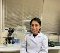
Graduate School of Medical and Dental Sciences Oral Life Science Tissue Regeneration and Reconstruction Assistant Professor

Updated on 2025/12/25


博士(歯学) ( 2014.9 新潟大学 )
Pulp Biology
Oral Histology
Tissue Regeneration
Tooth Development
Oral Anatomy
Gross Anatomy
Craniofacial Developmental Biology
Hard tissue
Life Science / Regenerative dentistry and dental engineering
Life Science / Developmental dentistry
Life Science / Oral biological science
Life Science / Anatomy
Faculty of Dentistry, Niigata University – Niigata, Japan. Division of Anatomy and Cell Biology of the Hard Tissue
2022.3
Faculty of Dentistry, Niigata University - Niigata, Japan. Division of Anatomy and Cell Biology of the Hard Tissue
2021.5 - 2022.2
School of Dentistry, Niigata University - Niigata, Japan Division of Anatomy and Cell Biology of the Hard Tissue Visiting researcher
2019.8
Científica del Sur University - Lima, Peru. School of Stomatology Adjunct faculty / Associated researcher
2018.3 - 2021.5
Center of Dental Medicine, University of Zurich - Zurich, Switzerland. Institute of Oral Biology Visiting postdoctoral researcher
2017.9 - 2018.2
New York University College of Dentistry - New York, USA. Department of Basic Science and Craniofacial Biology Postdoctoral associate
2015.8 - 2017.2
School of Dentistry, University of Texas at Houston - Texas, USA. Center for Craniofacial Biology Postdoctoral researcher
2015.4 - 2015.7
Niigata University Institute of Medicine and Dentistry, Academic Assembly Assistant Professor
2022.3
Niigata University Tissue Regeneration and Reconstruction, Oral Life Science, Graduate School of Medical and Dental Sciences Assistant Professor
2022.3
Niigata University Faculty of Dentistry PhD. in Oral Life Sciences
2010.10 - 2014.9
Country: Japan
Los Andes Private University School of Stomatology Master of Science in Stomatology
2008.4 - 2010.3
Country: Peru
National University of San Marcos Faculty of Dentistry DDS
2001.4 - 2006.12
Country: Peru
International Association of Dental Traumatology (IADT)
2023
The Japanese Society for Regenerative Medicine
2022
Japanese Association for Oral Biology (JAOB)
2011
The Japanese Association of Anatomists (JAA)
2011
International Association for Dental Research (IADR)
2009
Stress Distribution in a Mandibular Kennedy class I Restored with Bilateral Implant-Assisted Removable Partial Denture: A Finite Element Analysis
Dagny Ochoa-Escate, Freddy Valdez-Jurado, Romel Watanabe, Martha Pineda Mejía, Edwin Antonio Córdova Huayanay, Maria Soledad Ventocilla Huasupoma, Marcos Herrera Cisneros, Giovanna Lujan Larreátegui, Angela Quispe-Salcedo, Doris Salcedo-Moncada, Jesús Julio Ochoa Tataje
2025.2
Pulpal Responses to Leukocyte- and Platelet-Rich Plasma Treatment in Mouse Models for Immediate and Intentionally Delayed Tooth Replantation
Angela Quispe-Salcedo, Kiyoko Suzuki-Barrera, Mauricio Zapata-Sifuentes, Taisuke Watanabe, Tomoyuki Kawase, Hayato Ohshima
Applied Sciences 2024.12
マウス歯の再植時の意図的穿孔形成がマクロファージの時空間ダイナミクスに与える影響
佐野 拓人, 大島 邦子, Quispe-Salcedo A., 岡田 康男, 佐藤 拓一, 大島 勇人
Journal of Oral Biosciences Supplement 2024 [P1 - 13] 2024.11
Bases moleculares de la patogénesis del ameloblastoma: una revisión
Grecia Lourdes Riofrio Chung, Tania Lisseth Santos Tucto, Angela Quispe-Salcedo
Revista Científica Odontológica 2024.9
Effects of Synthetic Toll-Like Receptor 9 Ligand Molecules on Pulpal Immunomodulatory Response and Repair after Injuries. International journal
Angela Quispe-Salcedo, Tomohiko Yamazaki, Hayato Ohshima
Biomolecules 14 ( 8 ) 2024.8
Effects on dentin nanomechanical properties, cell viability and dentin wettability of a novel plant-derived biomodification monomer. International journal
Mário A Moreira, Madiana M Moreira, Diego Lomonaco, Eduardo Cáceres, Lukasz Witek, Paulo G Coelho, Emi Shimizu, Angela Quispe-Salcedo, Victor P Feitosa
Dental materials : official publication of the Academy of Dental Materials 2024.7
Early revascularization activates quiescent dental pulp stem cells following tooth replantation in mice International journal
Hiroto Sano, Kuniko Nakakura-Ohshima, Angela Quispe-Salcedo, Yasuo Okada, Takuichi Sato, Hayato Ohshima
Regenerative Therapy 24 582 - 591 2023.12
Mujeres en ciencia: Los desafíos de las dentistas-científicas en Perú
Angela Quispe-Salcedo
Odontología Sanmarquina 26 ( 3 ) 2023.9
Importancia de la técnica de perfusión intracardiaca para los estudios in vivo en Odontología
Mauricio Andre Zapata-Sifuentes, Kiyoko Suzuki-Barrera, Angela Quispe-Salcedo
Revista Científica Odontológica 2023.3
Explorando nuevos horizontes mediante la promoción de Programas de rotación de pregrado entre estudiantes de Odontología
Angela Quispe-Salcedo
Revista Científica Odontológica 2023.3
[Importance of intracardiac perfusion fixation technique for in vivo studies in dentistry]. International journal
Mauricio Andre Zapata-Sifuentes, Kiyoko Suzuki-Barrera, Angela Quispe-Salcedo
Revista cientifica odontologica (Universidad Cientifica del Sur) 11 ( 1 ) e148 2023
Perspectives on peer-review and editorial activities of Peruvian dental researchers. International journal
Angela Quispe-Salcedo
Revista cientifica odontologica (Universidad Cientifica del Sur) 11 ( 4 ) e170 2023
Actividad inhibitoria del extracto etanólico del Cyperus Rotundus procedente de la región de Cajamarca (provincia de Contumazá) en una cepa estandarizada de Streptococcus mutans (ATCC®25175) International journal
Rosita Belén Bazán Aliaga, Óscar Reátegui Arévalo, Luz Verónica Solórzano Espinoza, Juan Antonio Castro Arredondo, Víctor Elmo Miranda García, Elba Estefanía Martínez Cadillo, Angela Quispe-Salcedo
Revista Científica Odontológica 10 ( 1 ) e093 2022.4
The Critical Role of MMP13 in Regulating Tooth Development and Reactionary Dentinogenesis Repair Through the Wnt Signaling Pathway International journal
Henry F. Duncan, Yoshifumi Kobayashi, Yukako Yamauchi, Angela Quispe-Salcedo, Zhi Chao Feng, Jia Huang, Nicola C. Partridge, Teruyo Nakatani, Jeanine D’Armiento, Emi Shimizu
Frontiers in Cell and Developmental Biology 10 883266 - 883266 2022.4
Exploration of the role of the subodontoblastic layer in odontoblast-like cell differentiation after tooth drilling using Nestin-enhanced green fluorescent protein transgenic mice International journal
Chihiro Imai, Hiroto Sano, Angela Quispe-Salcedo, Kotaro Saito, Mitsushiro Nakatomi, Hiroko Ida-Yonemochi, Hideyuki Okano, Hayato Ohshima
Journal of Oral Biosciences 64 ( 1 ) 77 - 84 2022.3
Grupos de investigación: juntos llegamos lejos
Angela Quispe-Salcedo
Revista Científica Odontológica 2021.10
The effects of reducing the root length by apicoectomy on dental pulp revascularization following tooth replantation in mice International journal
Kuniko Nakakura-Ohshima, Angela Quispe-Salcedo, Hiroto Sano, Haruaki Hayasaki, Hayato Ohshima
DENTAL TRAUMATOLOGY 37 ( 5 ) 677 - 690 2021.10
Ezh2 knockout in mesenchymal cells causes enamel hyper- mineralization International journal
Yoshifumi Kobayashi, Angela Quispe-Salcedo, Sanika Bodas, Satoko Matsumura, Erhao Li, Richard Johnson, Marwa Choudhury, Daniel H. Fine, Siva Nadimpalli, Henry F. Duncan, Amel Dudakovic, Andre J. van Wijnen, Emi Shimizu
BIOCHEMICAL AND BIOPHYSICAL RESEARCH COMMUNICATIONS 567 72 - 78 2021.8
The Role of Dendritic Cells during Physiological and Pathological Dentinogenesis International journal
Angela Quispe-Salcedo, Hayato Ohshima
JOURNAL OF CLINICAL MEDICINE 10 ( 15 ) 2021.8
[Research groups: together we reach further]. International journal
Angela Quispe-Salcedo
Revista cientifica odontologica (Universidad Cientifica del Sur) 9 ( 3 ) e066 2021
[Basic dental sciences as the foundations for clinical practice]. International journal
Angela Quispe-Salcedo
Revista cientifica odontologica (Universidad Cientifica del Sur) 9 ( 2 ) e053 2021
An overview of Peruvian dental research in time of COVID-19
Angela Quispe-Salcedo
Revista Científica Odontológica 2020.12
Responses of oral-microflora-exposed dental pulp to capping with a triple antibiotic paste or calcium hydroxide cement in mouse molars International journal
Angela Quispe-Salcedo, Takuichi Sato, Junko Matsuyama, Hiroko Ida-Yonemochi, Hayato Ohshima
REGENERATIVE THERAPY 15 216 - 225 2020.12
Promotion of education and research in dental basic science. A call for action
Vilma Chuquihuaccha-Granda, Angela Quispe-Salcedo
Journal of Oral Research 9 ( 6 ) 446 - 448 2020.11
COVID-19 y su impacto en la odontología peruana
Angela Quispe-Salcedo
Revista Científica Odontológica 8 ( 1 ) 1 - 2 2020.4
Estudio in vitro del Efecto Antibacteriano de la Oleorresina de Copaifera reticulata y el Aceite Esencial de Origanum majoricum Frente a Streptococcus mutans y Enterococcus Faecalis Bacterias de Importancia en Patologías Orales
Angela Quispe-Salcedo
International journal of odontostomatology 2018.12
La importancia de las ciencias básicas en la formación del cirujano dentista
Angela Quispe-Salcedo
Odontología Sanmarquina 2018.9
Nestin expression is differently regulated between odontoblasts and the subodontoblastic layer in mice International journal
Mitsushiro Nakatomi, Angela Quispe-Salcedo, Masaka Sakaguchi, Hiroko Ida-Yonemochi, Hideyuki Okano, Hayato Ohshima
HISTOCHEMISTRY AND CELL BIOLOGY 149 ( 4 ) 383 - 391 2018.4
IGF-1 Mediates EphrinBl Activation in Regulating Tertiary Dentin Formation
S. Matsumura, A. Quispe-Salcedo, C. M. Schiller, J. S. Shin, B. M. Locke, S. Yakar, E. Shimizu
Journal of Dental Research 96 ( 10 ) 1153 - 1161 2017.9
Intercellular Genetic Interaction Between Irf6 and Twist1 during Craniofacial Development International journal
Walid D. Fakhouri, Kareem Metwalli, Ali Naji, Sarah Bakhiet, Angela Quispe-Salcedo, Larissa Nitschke, Youssef A. Kousa, Brian C. Schutte
SCIENTIFIC REPORTS 7 ( 1 ) 7129 - 7129 2017.8
The effects of enzymatically synthesized glycogen on the pulpal healing process of extracted teeth following intentionally delayed replantation in mice
Angela Quispe-Salcedo, Hiroko Ida-Yonemochi, Hayato Ohshima
JOURNAL OF ORAL BIOSCIENCES 57 ( 2 ) 124 - 130 2015.5
Effects of a Triple Antibiotic Solution on Pulpal Dynamics after Intentionally Delayed Tooth Replantation in Mice International journal
Angela Quispe-Salcedo, Hiroko Ida-Yonemochi, Hayato Ohshima
JOURNAL OF ENDODONTICS 40 ( 10 ) 1566 - 1572 2014.10
Use of a triple antibiotic solution affects the healing process of intentionally delayed replanted teeth in mice
Angela Quispe-Salcedo, Hiroko Ida-Yonemochi, Hayato Ohshima
JOURNAL OF ORAL BIOSCIENCES 55 ( 2 ) 91 - 100 2013.5
Expression patterns of nestin and dentin sialoprotein during dentinogenesis in mice
Angela Quispe-Salcedo, Hiroko Ida-Yonemochi, Mitsushiro Nakatomi, Hayato Ohshima
BIOMEDICAL RESEARCH-TOKYO 33 ( 2 ) 119 - 132 2012.4
EFFECT OF LEUKOCYTE PLATELET-RICH PLASMA ON OSSEOINTEGRATION OF IMPLANTS PLACED IN THE MOUSE MAXILLA
ZAPATA-SIFUENTES Mauricio, QUISPE-SALCEDO Angela, 渡辺泰典, 川瀬知之, 大島勇人
日本再生医療学会総会(Web) 23rd 2024
マウス顎骨へのインプラント埋入後のオッセオインテグレーションへのリンパ球-多血小板血漿(L-PRP)の効果
ZAPATASIFUENTES Mauricio Andre, QUISPE-SALCEDO Angela, WATANABE Taisuke, OHSHIMA Hayato
新潟歯学会雑誌 54 ( 1 ) 2024
Effectiveness of Leukocyte- and Platelet-Rich Plasma (L-PRP) on the pulpal healing process following tooth replantation in mice
QUISPE-SALCEDO Angela, ZAPATA-SIFUENTES Mauricio, WATANABE Taisuke, KAWASE Tomoyuki, OHSHIMA Hayato
Journal of Oral Biosciences Supplement (Web) 2023 2023
The positive effects of leukocyte- and platelet-rich plasma (l-prp) on osseointegration after implant placement in mouse maxilla
ZAPATA-SIFUENTES Mauricio Andre, QUISPE-SALCEDO Angela, WATANABE Taisuke, KAWASE Tomoyuki, OHSHIMA Hayato
Journal of Oral Biosciences Supplement (Web) 2023 2023
マウス臼歯再植後の早期血行回復は歯髄静的幹細胞を賦活化する
佐野拓人, 大島邦子, QUISPE-SALCEDO Angela, 岡田康男, 佐藤拓一, 大島勇人
Journal of Oral Biosciences Supplement (Web) 2023 2023
Effects of synthetic CpG oligodeoxynucleotides on the healing process of heavily injured tooth pulp
ANGELA Quispe-Salcedo, ANGELA Quispe-Salcedo, 山崎智彦, 依田浩子, 大島勇人
日本再生医療学会総会(Web) 22nd 2023
象牙芽細胞におけるNestin遺伝子の発現制御機構
中富 満城, Quispe-Salcedo Angela, 依田 浩子, 大島 勇人, 岡野 栄之
Journal of Oral Biosciences Supplement 2016 471 - 471 2016.9
象牙芽細胞におけるNestin遺伝子の発現制御機構
中富満城, QUISPE-SALCEDO Angela, 依田浩子, 大島勇人
Journal of Oral Biosciences Supplement (Web) 2016 2016
三種混合抗菌性薬剤と水酸化カルシウムセメント覆髄に対する感染歯髄の反応
Quispe-Salcedo Angela, 大島 勇人, 武藤 徳子, 石井 信之
神奈川歯学 50 ( 抄録集 ) 72 - 72 2015.11
酵素合成グリコーゲンによる歯の再植後の歯髄治癒促進効果について
大島勇人, QUISPE-SALCEDO Angela, 高田洋樹, 依田浩子
再生医療 13 2014
Root resection accelerates the dental pulp regeneration following tooth replantation in mice
QUISPE-SALCEDO Angela, IDA-YONEMOCHI Hiroko, OHSHIMA Hayato
日本解剖学会総会・全国学術集会講演プログラム・抄録集 119th 2014
離乳前後および成熟マウスの口腔内プラーク常在菌叢の網羅的解析
松山 順子, 佐藤 拓一, Quispe-Salcedo Angela, 高橋 信博, 大島 勇人
Journal of Oral Biosciences Supplement 2013 223 - 223 2013.9
マウスの意図的に遅延した歯の生え替わり後の治癒過程に対する酵素的に合成したグリコーゲン(ESG)の有効性(Effectiveness of Enzymatically Synthesized Glycogen (ESG) on the healing process following intentionally-delayed tooth replantation in mice)
Quispe-Salcedo Angela, 依田 浩子, 大島 勇人
Journal of Oral Biosciences Supplement 2013 123 - 123 2013.9
Effectiveness of Enzymatically Synthesized Glycogen(ESG)on the healing process following intentionally-delayed tooth replantation in mice
QUISPE-SALCEDO Angela, IDA Hiroko, OSHIMA Hayato
Journal of Oral Biosciences Supplement (Web) 2013 2013
離乳前後および成熟マウスの口腔内プラーク常在菌叢の網羅的解析
松山順子, 佐藤拓一, QUISPE-SALCEDO Angela, 高橋信博, 大島勇人
Journal of Oral Biosciences Supplement (Web) 2013 2013
意図的に遅延した歯の再植後の歯髄の治癒過程における抗菌薬の有効性(Effectiveness of antimicrobials in the pulpal healing process following intentionally delayed tooth replantation)
Quispe-Salcedo Angela, 依田 浩子, 大島 勇人
Journal of Oral Biosciences Supplement 2012 85 - 85 2012.9
マウス口腔内プラーク常在菌叢の網羅的解析
松山 順子, 佐藤 拓一, Quispe-Salcedo Angela, 石田 直子, 高橋 信博, 大島 勇人
Journal of Oral Biosciences Supplement 2012 138 - 138 2012.9
Effectiveness of antimicrobials in the pulpal healing process following intentionally delayed tooth replantation
QUISPE-SALCEDO Angela, IDA Hiroko, OSHIMA Hayato
Journal of Oral Biosciences Supplement (Web) 2012 2012
マウス口腔内プラーク常在菌叢の網羅的解析
松山順子, 佐藤拓一, ANGELA Quispe-Salcedo, 石田直子, 石田直子, 高橋信博, 大島勇人
Journal of Oral Biosciences Supplement (Web) 2012 2012
In vivo assessment of synthetic toll-like receptor 9 ligand molecules for the treatment of the afflicted dental pulp following tooth replantation in mice
Angela Quispe-Salcedo, Tomohiko Yamazaki, Hayato Ohshima
22nd World Congress on Dental Traumatology 2024, Tokyo, Japan 2024.7
Effects of synthetic toll-like receptor 9 ligand molecules on the pulpal immunomodulatory responses and repair after injuries
Angela Quispe-Salcedo, 山崎智彦, 大島勇人
129th Annual Meeting of the Japanese Association of Anatomists, Okinawa, Japan 2024.3
Avances en la reacción de la pulpa dental
Angela Quispe-Salcedo
Congreso Internacional de la Facultad de Odontologia de laUniversidad Latina, Costa Rica (online) 2023.10
Effectiveness of Leukocyte- and Platelet-Rich Plasma (L-PRP) on the pulpal healing process following tooth replantation in mice.
Quispe-Salcedo A, Zapata-Sifuentes M, Watanabe T, Kawase T, Ohshima H
The 65th Annual Meeting of the Japanese Association for Oral Biology. Tokyo, Japan. 2023.9
Effects of synthetic CpG oligodeoxynucleotides on the healing process of heavily injured tooth pulp.
Quispe-Salcedo A, Yamazaki T, Ohshima H
22nd Annual Meeting of the Japanese Society for Regenerative Medicine. Kyoto, Japan. 2023.3
The interaction between osteopontin and stem/progenitor cells determines the pulpal healing following tooth replantation in mice.
Quispe-Salcedo A, Suzuki-Barrera K, Nakatomi M, Ida-Yonemochi H, Ohshima H
The 64th Annual Meeting of the Japanese Association for Oral Biology. Tokushima, Japan. 2022.9
Effectiveness of 3Mix-MP Paste as Pulp-Capping Agent in Murine Molars.
Quispe-Salcedo A, Sato T, Matsuyama J, Ida-Yonemochi H, Ohshima H
IADR/ AADR / CADR General Session Washington DC, USA. 2020
Bases biológicas y evidencia científica del uso de la pasta triple antibiótica 3Mix en patologías pulpares.
Quispe-Salcedo A
VII Congreso de la Región Latinoamericana IADR. XIII Reunión Científica SUIO. Montevideo, Uruguay. 2018.8
Histone Methyltransferaase Ezh2 is Required for Enamel Maturation
Quispe-Salcedo A, Matsumura S, Li E, Dudakovic A, van Wijnen AJ, Shimizu E
IADR/ AADR / CADR General Session. San Francisco, California. USA. 2017.3
ephrinB1 ligand is a critical mediator for tertiary dentinogénesis.
Quispe-Salcedo A, Matsumura S, Schiller C, Yakar S, Shimizu E
IADR Pulp biology and regeneration group symposium. Nagoya, Japon. 2016.6
Responses of infected dental pulp to capping with the mixture of three antibacterial drugs (3Mix) or calcium hydroxide cement in mouse molars.
Quispe-Salcedo A, Sato T, Matsuyama J, Ohshima H
The 56th Annual meeting of the Japanese Association for Oral Biology. Fukuoka, Japan 2014.9
Root resection accelerates the dental pulp regeneration following tooth replantation in mice.
Quispe-Salcedo A, Ohshima H
The 119th Annual Meeting of the Japanese Association of Anatomists. Tochigi, Japan 2014.3
The resection of the root accelerates pulpal regeneration following tooth replantation in mice.
Quispe-Salcedo A, Ohshima H
International Symposium Frontier Meeting. Niigata, Japan 2014.2
Effectiveness of enzymatically synthesized glycogen (ESG) on the healing process following intentionally-delayed tooth replantation in mice.
Quispe-Salcedo A, Ida-Yonemochi H, Ohshima H
The 55th Annual meeting of the Japanese Association for Oral Biology. Okayama, Japan 2013.9
The use of antimicrobials accelerates the dental pulp regeneration following intentionally-delayed tooth replantation in mice.
Quispe-Salcedo A, Ida-Yonemochi H, Ohshima H
11th International Conference on Tooth Morphogenesis and Differentiation. La Londe les Maures, France 2013.6
Expression of GFP and nestin immunoreactivity during postnatal tooth development in nestin-EGFP transgenic mice.
Quispe-Salcedo A, Ida-Yonemochi H, Mitsushiro N, Nakagawa E, Saito K, Okano H, Ohshima H
The 118th Annual Meeting of the Japanese Association of Anatomists. Takamatsu, Japan 2013.3
The use of antimicrobials accelerates the pulpal healing process following intentionally-delayed tooth replantation in mice.
Quispe-Salcedo A, Ida-Yonemochi H, Ohshima H
International Symposium Frontier Meeting: Development, Evolution, Taxonomy, and Genetics of Tooth Structure “Tooth Voyage, Up To Date”. Jeonju, South Korea 2013.2
Effectiveness of antimicrobials in the pulpal healing process following intentionally delayed tooth replantation.
Quispe-Salcedo A, Ida-Yonemochi H, Ohshima H
The 54th annual meeting of the Japanese Association for Oral Biology. Koriyama, Japan 2012.9
The application of antimicrobials or glycogen accelerates the pulpal regeneration of replanted molars in mice. March 2012.
Quispe-Salcedo A, Ida-Yonemochi H, Ohshima H
The 117th Annual Meeting of the Japanese Association of Anatomists. Yamanashi, Japan 2012.3
Assessment of nestin and dentin sialoprotein expression patterns during dentinogenesis and aging.
Quispe-Salcedo A, Ida-Yonemochi H, Mitsushiro N, Ohshima H
The 53th Annual meeting of the Japanese Association for Oral Biology. Gifu, Japan 2011.9
Assessment of intermediate filament nestin and dentin sialoprotein expression patterns in the process of odontoblast differentiation.
Quispe-Salcedo A, Ida-Yonemochi H, Nakatomi M, Kenmotsu S, Ohshima H
The 116th Annual Meeting of the Japanese Association of Anatomists. Yokohama, Japan 2011.3
Postdoctoral Fellowship for Research in Japan
2021 Japan Society for the Promotion of Science (JSPS)
Travel Grant for Young New Investigators. 98th IADR General Session – Washington DC. USA.
2020 International Association for Dental Research (IADR)
ravel Grant for Short-Term Research Stay in Niigata University, Japan
2019 National Fund for Scientific Development, Technology and Technology Innovation (FONDECYT) – Peru
STAR Network Academy Fellowship
2019 International Association for Dental Research (IADR)
Swiss Government Excellence Scholarship for postdoctoral studies
2017 Federal Commission for Scholarships (FCS)
Best paper award in the Journal of Oral Biosciences - 2015
2015 Japanese Association for Oral Biology
Japanese government (Monbukagakusho) scholarship for research students.
2010 Japanese Ministry of Education, Culture, Sports, Science and Technology
Homeostatic maintenance and activation of dental pulp quiescent stem/progenitor cells regulated by dendritic cells and macrophages
Grant number:23H03078
2023.4 - 2026.3
System name:Grants-in-Aid for Scientific Research
Research category:Grant-in-Aid for Scientific Research (B)
Awarding organization:Japan Society for the Promotion of Science
Grant amount:\18980000 ( Direct Cost: \14600000 、 Indirect Cost:\4380000 )
Cross-talk among odontoblasts, dental pulp stem cells, and immune cells after exogenous injuries
Grant number:22K21011
2022.8 - 2024.3
System name:Grants-in-Aid for Scientific Research
Research category:Grant-in-Aid for Research Activity Start-up
Awarding organization:Japan Society for the Promotion of Science
Grant amount:\2860000 ( Direct Cost: \2200000 、 Indirect Cost:\660000 )
Cross-talk among odontoblasts, dental pulp stem cells, and immune cells after exogenous injuries
Grant number:21F30412
2021.7 - 2022.3
System name:Grants-in-Aid for Scientific Research
Research category:Grant-in-Aid for JSPS Fellows
Awarding organization:Japan Society for the Promotion of Science
Grant amount:\2300000 ( Direct Cost: \2300000 )
外的侵襲後の歯髄治癒過程における象牙芽細胞、歯髄幹細胞、免疫細胞間クロストーク
Grant number:21F20412
2021.4 - 2023.3
System name:科学研究費助成事業
Research category:特別研究員奨励費
Awarding organization:日本学術振興会
大島 勇人, QUISPE SALCEDO ANGELA
Grant amount:\2300000 ( Direct Cost: \2300000 )
Osteology
Gross Anatomy
General and Oral Embryology
General and Oral Histology
人体解剖学I
人体解剖学実習
人体解剖学II
Vice-president
Role(s): Presenter
International Association for Dental Research – Peruvian Division 2019 - 2021
Science Clubs Peru (Clubes de Ciencia Peru)
Role(s): Presenter
2017
Associate Editor
Revista Cientifica Odontologica (Lima) 2018
Review Editor
Role(s): Peer review
Frontiers in Physiology 2017
Review Editor
Role(s): Peer review
Frontiers in Dental Medicine